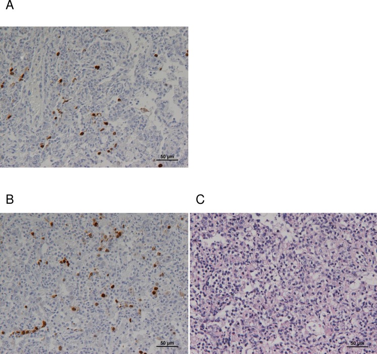
Fig 3. Detection of viral antigen in lung sections.
Sections from (A) upper left and (B) lower left lung from one NHP (i.t. challenge 5 days post-infection) were stained with monoclonal antibody to IAV nucleoprotein and visualised by the PAP method (brown staining). Panel (C) shows an H&E stained section from the same sample as (B).

