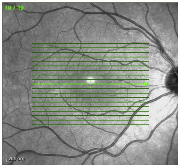
FIG 2.
Three macular regions selected for analysis: superior macula, central macula including presumed central fovea, and inferior macula. “Central” sections represent segmentation through the presumed foveal center as well as one section 250 μm inferior and superior to the foveal center. “Superior” and “inferior” sections delineate 2 segments of retina 1250 μm and 1500 μm superior or inferior to the foveal center, respectively.

