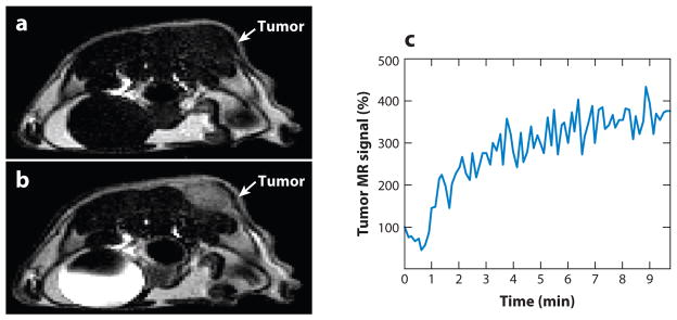
Figure 1.
Contrast enhancement of pathological tissues with a T1 magnetic resonance imaging (MRI) contrast agent. (a) The tumor of an MDA-MB-231 mammary carcinoma model is difficult to identify without the MRI contrast agent. (b) T1 contrast agent Gd-DTPA (Magnevist™) accumulated in the tumor and enhanced the image contrast for the tumor. (c) The temporal change in the T1-weighted MR signal of the tumor confirmed that the contrast enhancement was due to accumulation of the agent. Reproduced with permission from Reference 9.

