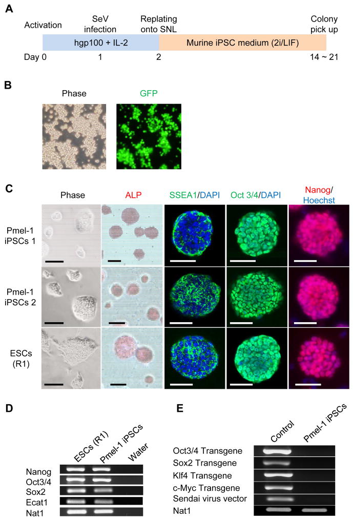
Figure 1. Generation of iPSCs from Pmel-1 TCR transgenic CD8+ T cells.
(A) Schematic illustration showing the generation of iPSCs from Pmel-1 TCR transgenic CD8+ T cells. (B) Efficient GFP introduction by Sendai virus vector in Pmel-1 CD8+ T cells transfected at an MOI of 20. (C) Morphology, alkaline phosphatase (ALP) activity and expression of pluripotency and surface markers (SSEA1, Oct3/4 and Nanog) in Pmel-1 iPSCs 1 and 2, and mESCs. Nuclei were counterstained with DAPI or HOECHST. Scale bar: 200 μm. (D) RT-PCR analysis for the ESC marker genes, Nanog, Oct3/4, Sox2, and Ecat1 in Pmel-1 iPSCs and mESCs. (E) RT-PCR analysis for the transgenes, Oct3/4 Sox2, Klf4, c-Myc and Sendai virus in Pmel-1 iPSCs.

