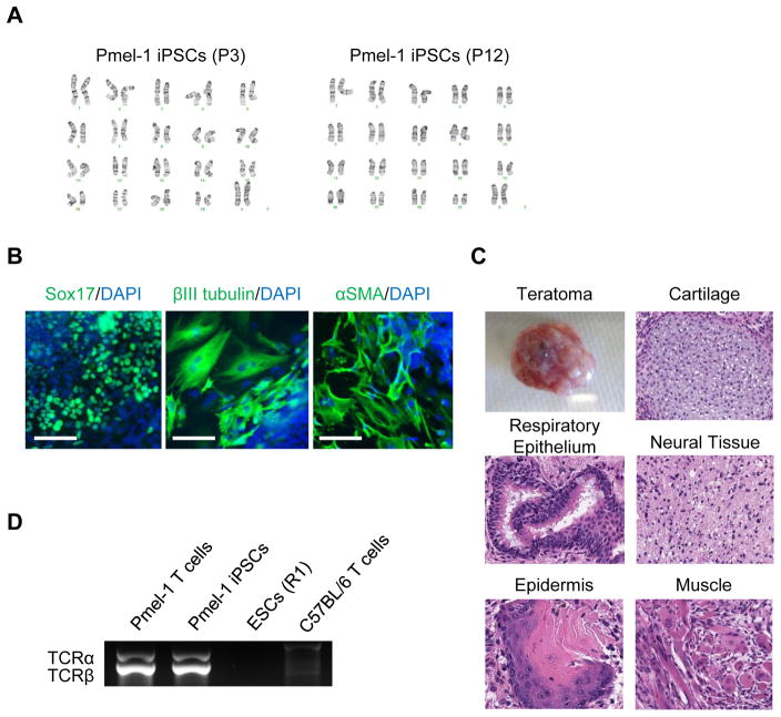
Figure 2. Detail characterizations of Pmel-1 CD8+ T cell-derived iPSCs.
(A) Normal karyogram of the Pmel-1 iPSCs at passage 3 (left) and 12 (right). (B) Immunostaining for Sox17 (endodermal marker), βIII tubulin (ectodermal marker), and αSMA (mesodermal marker) in Pmel-1 iPSC-derived differentiated cell. Scale bar: 100 μm (C) Gross morphology, hematoxylin and eosin-stained representative teratomas derived from Pmel-1 iPSCs. (D) PCR analysis of rearranged Pmel-1 TCR α- and β-chain genes in Pmel-1 T cells, Pmel-1 iPSCs, mESCs and C57BL/6 mouse T cells.

