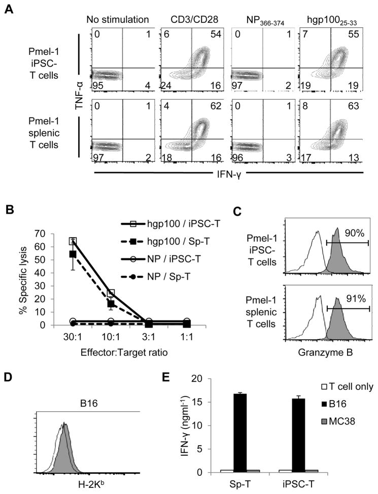
Figure 4. iPSC-derived CD8+ T Cells exhibit antigen-specific cytokine production and cytolysis in vitro.
(A) Intracellular production of IFN-γ and/or TNF-α by Pmel-1 iPSC-derived and splenic CD8+ T cells upon stimulation with or without anti-CD3/CD28 mAb, or against the hgp10025-33 or NP366-374 antigens. CD8-gated populations were shown. Numbers denote percent positive cells. (B) Cytolytic function of Pmel-1 iPSC-derived and splenic T cells against the hgp10025-33 or NP366-374 antigens is shown. (C) Intracellular production of granzyme B induced by stimulation of Pmel-1iPSC-derived and splenic CD8+ T cells with hgp100 pulsed MC38 tumor cells. (D) MHC-I (H-2Kb) expression on B16 melanoma tumors. Open histograms in (C) and (D) indicate the isotype-control staining. (E) Production of IFN-γ by Pmel-1 iPSC-derived and splenic T cells against B16 melanomas and MC38 colon adenocarcinomas. Data shown in (A) – (E) are representative of 2 independent experiments.

