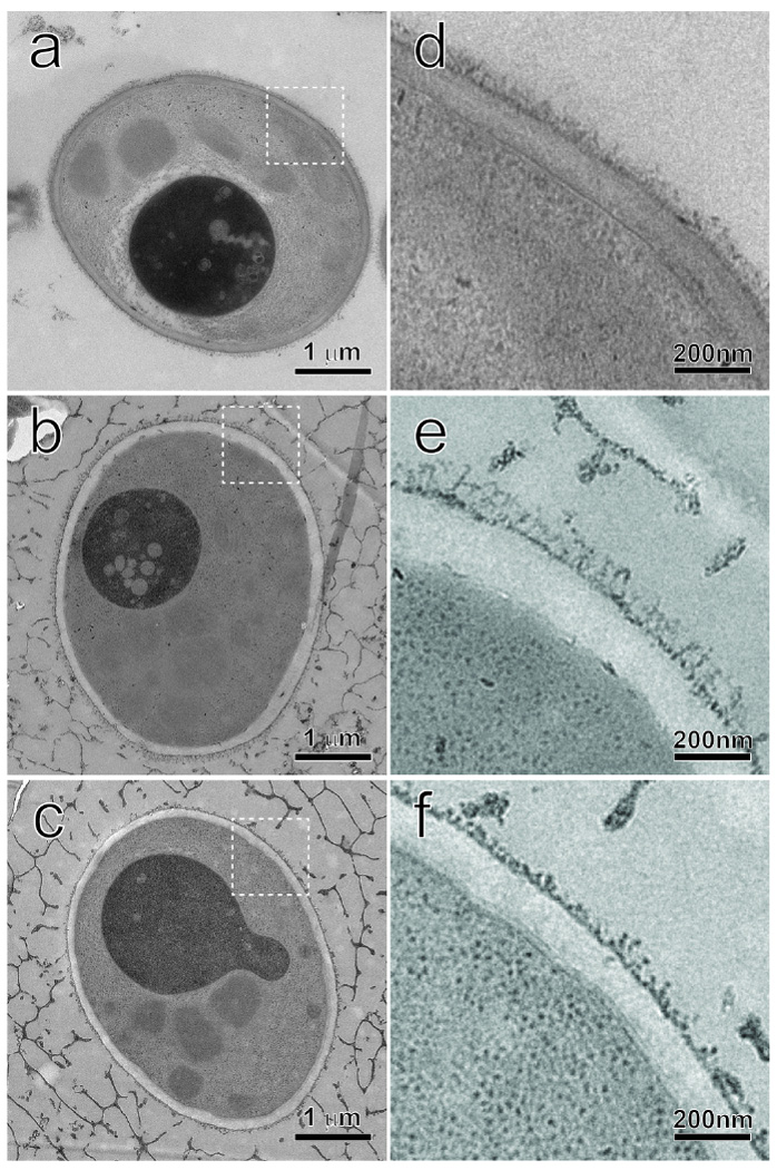
Fig.9.

Electron microscopy of the three recombinant yeast strains. The cells were rapidly frozen after methanol induction for 96 h. Following freeze-substitution, infiltration and polymerization, the sample blocks were serially sectioned to a thickness of about 100 nm. The structural preservation is very good as demonstrated in the whole-cell micrographs in a GS115/CALB-GCW51, b GS115/SC3-61/CALB-51, and c GS115-51/HFBI-61/CALB-51. The cell wall regions, marked by the dash-line squares in the left figure column, are further magnified in d GS115/CALB-GCW51, e GS115/SC3-61/CALB-51 and f GS115-51/HFBI-61/CALB-51, respectively. The difference of the inner layers and the outer mannan fibrils are visible among the three strains.
