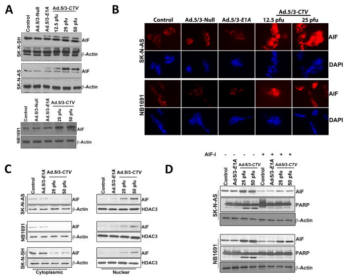
Figure 4. Ad.5/3-CTV promotes AIF-mediated cell death in neuroblastoma cells.
(A) Neuroblastoma cells were infected with 25 pfu of Ad.5/3-Null or Ad.5/3-E1A or with the indicated dose of Ad.5/3-CTV for 72 hours. Cells were collected and western blotting analysis was performed for AIF using specific antibodies and β-Actin served as loading control. Results are representative of three independent experiments. (B) Neuroblastoma cells were cultured in 8-well chamber slide and treated as described in 4A for 72 hours. These cells were then subjected to immunofluorescence analysis of AIF using anti-AIF antibody and Alexa Flour-594 secondary antibody (red fluorescence). Nuclei were stained with DAPI (blue fluorescence). Fluorescent cells were visualized and photographed from 10 different fields and representative images are shown. (C) Subcellular distribution of AIF was determined using western blot analysis. Neuroblastoma cells were infected with 25 pfu of Ad.5/3-E1A or the indicated dose of Ad.5/3-CTV for 72 hours. The cytoplasmic and nuclear fractions were isolated and examined by western blotting for AIF using specific antibodies. HDAC3 served as loading control for nuclear extract and β-Actin served as loading control for cytoplasmic extracts. (D) Neuroblastoma cells were pre-treated with AIF inhibitor and infected with 25 pfu of Ad.5/3-E1A or the indicated dose of Ad.5/3-CTV for 48 hours. Cells were collected and western blotting analysis was performed for AIF and PARP using specific antibodies and β-Actin served as loading control. Results are representative of three independent experiments.

