
Figure 1.
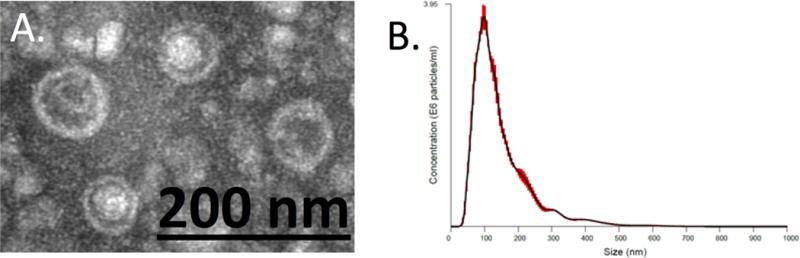
Characterization of LA-derived EVs. (A) Transmission electron microscopy (TEM) analysis of EVs obtained by ultracentrifugation showing spherical structures enclosed by a lipid bilayer. (B) Nanoparticle tracking analysis (NTA) of colonic EV obtained by ultracentrifugation. Distribution of EV particle concentration (y axis) by size (x axis). Red area is the standard error.