
Figure 2.
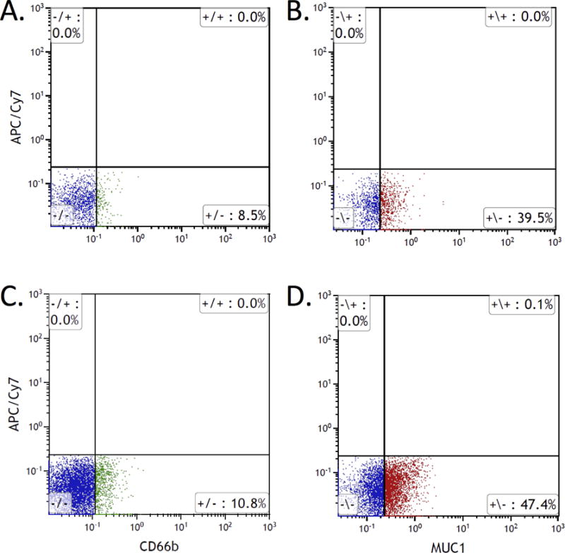
Flow cytometry analysis of EVs isolated from colonic luminal aspirates. Purified EVs from HC (A & B) and IBD patients (C & D) were stained with anti-CD63 combined with either anti-CD66b (A & C) or anti-MUC-1 (B & D). All images are the gating of CD63 positive particles within EV gate (see Supplementary Fig. 2).