
Figure 6.
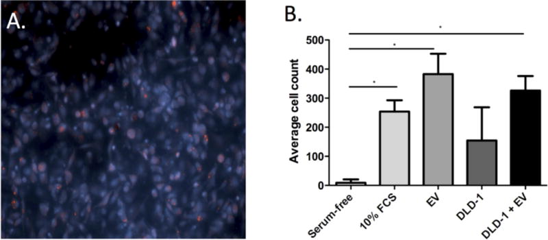
Macrophage migration assay. Images of marcophages on trans-well membranes after exposure of macrophages to EVs from IBD patients labelled with acridine orange (red), and the nucleic acids of macrophages were stained with Hoechst 33258 (blue) (A, 20× magnification); (B) Mean cell counts on the trans-well membrane after 10 hours of exposure of macrophages to serum-free medium (negative control), 10% FBS (positive control), EVs from IBD patients (IBD EV), DLD-1 cells alone, or DLD-1 cells cultured with IBD EVs. Note that error bars are standard deviations.