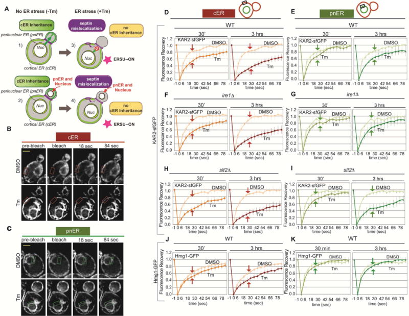
Figure 1. ER stress has a greater effect on Kar2-sfGFP mobility in the cER than in the pnER.

(A) Illustration of the effects of ER stress on tubule formation and cER and pnER inheritance. Under unstressed conditions, an ER tubule formed from the pnER moves from the mother into the bud where it forms the cER (1). This is followed by nuclear migration (2). Under conditions of ER stress, tubule formation is abnormal and the cER is not inherited (3), generating buds that contain the pnER but no cER (4).
(B and C) Representative image of WT cells expressing Kar2-sfGFP. FRAP analysis monitored Kar2-sfGFP mobility in the cER (B) or pnER (C) of DMSO or Tm (1 μg/ml, 3hr) treated cells. Images were acquired before (pre-bleach), at the same time as (bleach), and at 18 or 84 sec after photobleaching. The boxed area shows the cER (orange box) and pnER (green box) photobleached. Scale bar is 2 μm.
(D and E) Fluorescence intensity was normalized to the pre-bleach signal and recovery was plotted over time for the cER (D) and pnER (E) of WT cells treated with DMSO or Tm for 30 min or 3 hr. Graphs are the mean ± SD of 3 experiments, each examining ≥7 cells.
(F and G) Kar2-sfGFP mobility in the cER and pnER of ire1Δ cells is similar to WT cells. (H and I) slt2 deletion reduces Kar2-sfGFP mobility in the cER and pnER of stressed cells. (J and K) As described for D and E except the experiments were performed with WT cells expressing Hmg1-GFP. Experiments in F–K were performed as in D and E. See also Figures S1 and S2.
