Fig. 1.
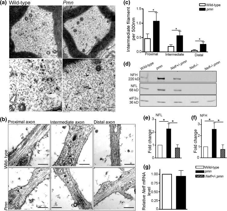
Analysis of intermediate filament (IF) levels in axons of pmn mutant mouse motoneurons. a Electron micrograph of cross sections of distal phrenic nerve from 34-day-old mice showed increased intermediate filaments in pmn axons compared to wild-type axons, scale bar 500 nm (top). Bottom lane shows the higher magnification of the respective image in the top lane (scale bar 200 nm). Arrows point to IF. b Electron micrograph showing axonal compartments of motoneurons cultured for 7 days. Scale bar 500 nm. c Quantification of number of IF in pmn axons showed an increase in proximal (P value = 0.0304), intermediate (P value = 0.0249) and distal (P value = 0.0336) compartments as compared to wild-type axons (Mann–Whitney one tailed test, n = 16 wild-type motoneurons and 19 pmn motoneurons from 3 independent experiments). d Western blot analysis of sciatic nerve lysate from 34-day-old mice. e, f Pmn mutant mouse nerves show increased levels of e NFL (t = 3.210, P = 0.0326) and f NFH (t = 2.781, P = 0.0498) proteins as compared to the wild type. Bars represent mean ± SEM, (n = 5 wild-type and pmn and n = 3 Nefl+/−;pmn mice analyzed, *P < 0.05; one sample t test). g Expression levels of Nefl mRNA in sciatic nerve extracts of 28- to 30-day-old pmn is not changed compared to wild type. Quantification was performed by normalizing with HPRT1 as housekeeping gene. Bars represent mean ± SEM (n = 3)

