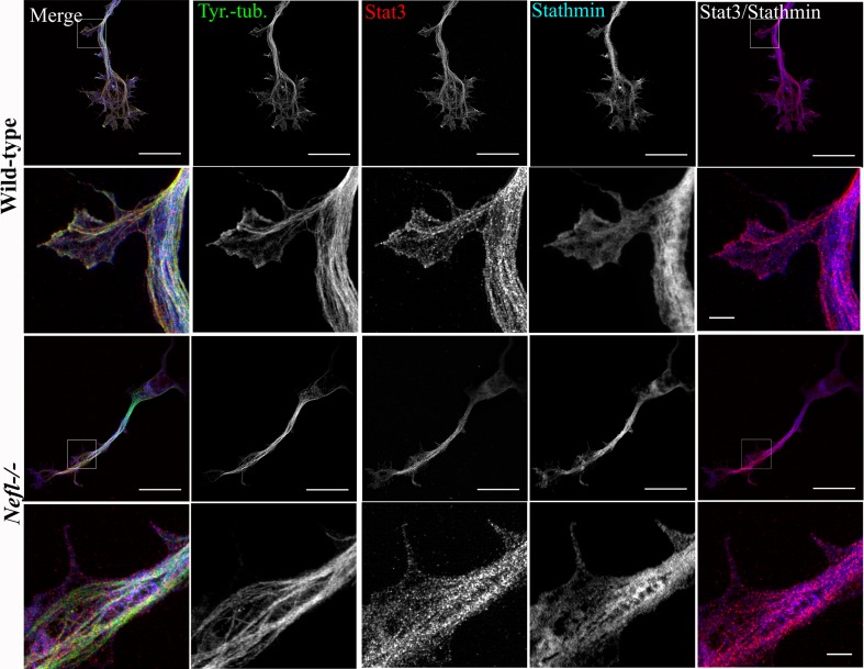
Fig. 7.
Distribution of Stat3 and stathmin in axons of wild-type and Nefl−/− motoneurons as revealed by high-resolution SIM. Motoneurons were cultured for 3 days in vitro. Representative images of wild-type and Nefl−/− motoneurons, stained with antibodies against tyrosinated α-tubulin (green), Stat3 (red), and stathmin (blue). The antibody against tyrosinated α-tubulin stains both soluble αβ-tubulin heterodimers and polymerized highly dynamic microtubules. Note that the colocalization of stathmin with Stat3 increases in Nefl−/− motoneurons (right panel). Scale bar 20 µm (first and third lane). White square boxes indicate the regions enlarged in the second and fourth lane, scale bar 2 µm

