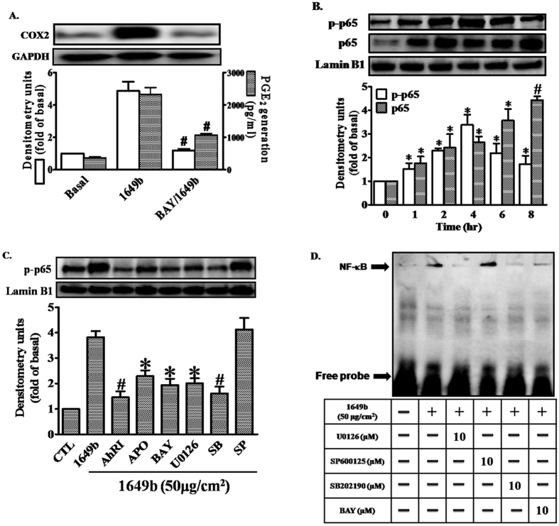
Figure 5. PMs induce COX2 expression via Nox2/ERK/p38/NF-κB signaling.
Cells were pre-incubated with NF-κB inhibitor (Bay11-7082; BAY) (10 μM) for 1 h and then exposed to PMs (50 μg/cm2) for 24 h. (A) COX2 protein expression (open bars) and prostaglandin E2 (PGE2) production (shaded bars) were determined by Western blotting and ELISA as described in Fig. 1. (B,C) Cells were pre-incubated with with AhRI (10 μM), APO (100 μM), U0126 (10 μM), SB202190 (10 μM), SP60015 (10 μM) or BAY (10 μM) for 1 h and then exposed to PMs (50 μg/cm2) for the 2 h. The levels of p65 in nuclear extract (detected with p65 or phospho-p65 antibodies), determined by Western blotting (C). Lamin-B1 was used as the loading control. (D) Cells were pre-incubated with with U0126 (10 μM), SB202190 (10 μM), SP60015 (10 μM) or BAY (10 μM) for 1 h and then exposed to PMs (50 μg/cm2) for 2 h. Electrophoretic mobility-shift assay for assessment of NF-κB DNA binding activity, as described in “Materials and methods”. (A–D) Data are expressed as mean ± standard error of the mean, based on three independent experiments. *P < 0.05, #P < 0.01, compared with basal levels.

