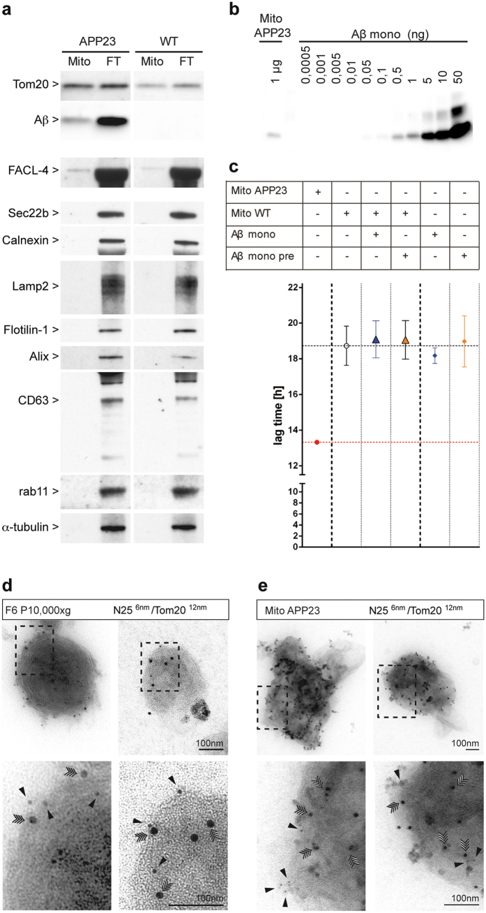
Figure 2. Immunoisolated mitochondria and associated ER-membranes contain Aβ and exhibit in-vitro seeding activity.
(a) Immunoisolated mitochondria and the corresponding flowthrough were prepared from amyloid-laden transgenic APP23 (n = 3) and old wild-type C57BL/6 mice (n = 3) and analyzed by immunoblotting for Aβ peptide (monoclonal Aβ 6E10), and markers for mitochondria (Tom20 and FACL4) endoplasmic reticulum (sec22b, Calnexin), lysosomes/late endosomes (Lamp2), lipid rafts (Flotilin-1), exosomes (Alix, CD63 and Rab11) and cytoskeletal protein α-tubulin. Note that full-length blots/gels are presented in Supplementary Fig. 6. (b) Estimation of the Aβ amount associated with 1 μg isolated mitochondria from APP23 mice by comparison with increasing amounts of Aβ(1–40) (c) in-vitro FRANK assay of APP23 and WT mitochondria (1 μg total protein). Where indicated WT mitochondria were spiked with 1 ng of monomeric Aβ(1–40) and either tested immediately or preincubated for 30 minutes at 37 °C. Monomeric Aβ(1–40) without mitochondria was used as control. Overview of the negative staining of (d) membrane vesicles from P10,000 × g fraction 6 and of (e) mitochondria immunoisolated from cortex of depositing APP23 mice after co-immunogold labeling for Aβ peptides with monoclonal JRF/AbN/25 antibody (against the free amino terminus and the first seven amino acids of human Aβ(1–40/42) peptides)(N25) and for mitochondria using a Tom20 antibody. Two examples of each preparation show the double-labeled membrane particles adsorbed on EM grids without sectioning. The boxed region indicates the area shown at higher magnification underneath. Note the double immunogold labeling on the membrane surface for Aβ peptides (N25, 6 nm gold, black arrowheads) and Tom20 (12 nm gold, quadruple-headed arrowheads). Bars: 100 nm. Note that full-length blots/gels are presented in Supplementary Fig. 7.

