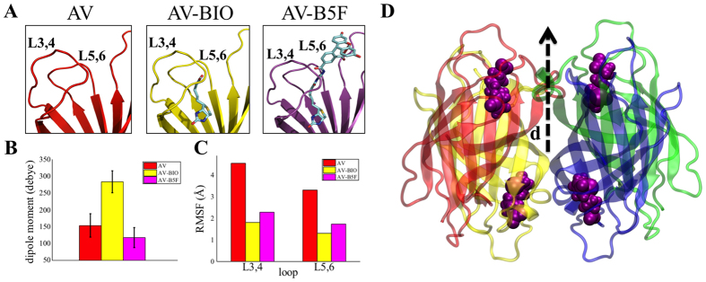
Figure 4. Molecular dynamics simulations of Biotin-binding proteins.
(A) Residues close to the binding pocket of the AV (left), AV-Biotin complex (center) and Avidin-B5F complex (right). (B) Histogram showing the dipole moment (Debye) averaged along the obtained molecular dynamics trajectory for AV (red), AV-Biotin (yellow) and AV-B4F (magenta) systems. A single value is reported for each system averaging statistics from the two monomers. (C) Histogram showing the computed Root Mean Square Fluctuations (RMSF) (Å) values for AV (red), AV-BIO (yellow) and AV-B4F (magenta) systems considering both L3,4 and L5,6 loops. For each system, the averaged RMSF, computed from the two monomers, is reported. (D) Overall tetrameric structure of the simulated AV with bound biotin molecules. Dipole directions are shown (not to scale).

