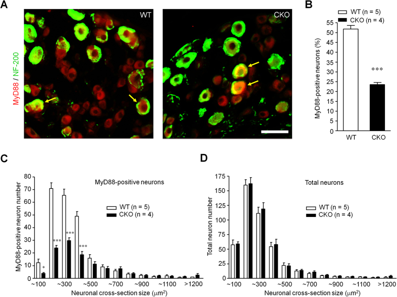
Figure 1. Deletion of Myd88 adapter protein in small-sized DRG neurons of CKO mice.
(A) Double immunostaining showing MyD88 localization in DRG neurons of littermate control (WT) and MyD88 conditional knockout (CKO) mice. Arrows indicate neurons with co-localization of MyD88 and NF200. Scale, 50 μm. (B) Percentage of MyD88-positve neurons in WT and MyD88 CKO mice. Six DRG sections were included per mouse. ***P < 0.001, two-tailed unpaired student T-test compared with WT; n = 4~5 mice/group. (C,D) Size distribution of MyD88-positive neurons (C) and total neurons (D) in WT and MyD88 CKO mice. Six DRG sections were counted per mouse. *P < 0.05, ***P < 0.001, two-way ANOVA compared with WT; n = 4~5 mice/group.

