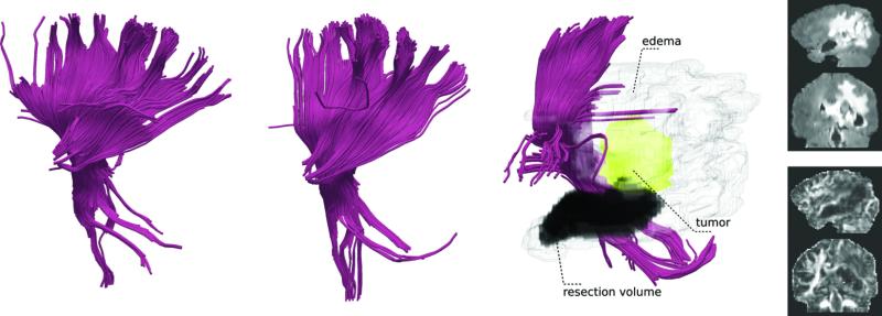
Figure 4.
Illustration of the WM fibers in the internal capsule in the atlas (first), a healthy participant (second), and a patient with a brain tumor and a prior surgical site (third). Surrounding edema around the tumor and resection volume is also shown. The part of the internal capsule that was reconstructed by the tractography was successfully identified by adaptive clustering in both healthy participant and the patient with the tumor despite the presence of large mass effect.

