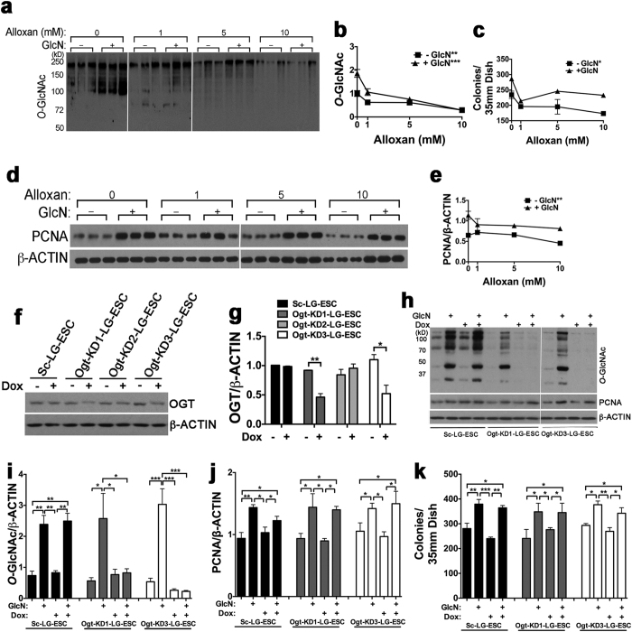
Figure 3. Effects of O-GlcNAcylation inhibition on LG-ESC proliferation.
(a) Immunoblot of total O-GlcNAcylated proteins from LG-ESC grown ± 0.8 mM GlcN and 0, 1, 5 or 10 mM alloxan. (b) Quantitation of (a). (c) Quantitation of AP+ LG-ESC colonies following culture as in a. (d) Immunoblot of PCNA and β-ACTIN following culture as in (a). (e) Quantitation of (d). (f) Immunoblot of O-GlcNAc transferase (OGT) from clones of LG-ESC stably transfected with doxycycline (Dox)-inducible pSingle containing a scrambled shRNA sequence (Sc-LG-ESC) or one of three different shRNA sequence targeting Ogt mRNA (Ogt-KD1-, Ogt-KD2-, or Ogt-KD3-LG-ESC), cultured ± 1 μg/ml Dox. (g) Quantitation of OGT/β-ACTIN assayed as in (f). (h) Immunoblot of total O-GlcNAcylated proteins, PCNA, and β-ACTIN from Sc-LG-ESC, Ogt-KD1-LG-ESC, or Ogt-KD3-LG-ESC treated ± 1 μg/ml Dox and ± 0.8 mM GlcN. (i) Quantitation of O-GlcNAcylated proteins from Sc-LG-ESC, Ogt-KD1-LG-ESC, or Ogt-KD3-LG-ESC assayed as in (h). (j) Quantitation of PCNA from Sc-LG-ESC, Ogt-KD1-LG-ESC, or Ogt-KD3-LG-ESC assayed as in (h). (k) Quantitation of AP+ colonies of Sc-LG-ESC, Ogt-KD1-LG-ESC, or Ogt-KD3-LG-ESC cultured ± 1 μg/ml Dox and ± 0.8 mM GlcN. Experiments were repeated 2–3 times using triplicate culture dishes. Data from representative experiments are displayed as the mean ± s.e.m. (N = 3) and were analyzed by linear regression (b,c, and e), Student’s t test (g) or two-way ANOVA followed by Tukey’s post test of each clone (i–k). *P < 0.05, **P < 0.01, ***P < 0.001.

