Figure 6.

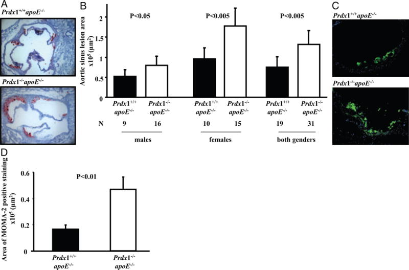
Atherosclerosis is accelerated in Prdx1−/−/apoE−/− mice. Animals were fed a normal chow diet and evaluated for aortic sinus lesions at 16 weeks of age. A, Atherosclerotic lesions were measured by lipid deposition detected with oil red O staining, which is represented here in red within aortic sinus. Representative images showing plaque (red) in female Prdx1−/−/apoE−/− and Prdx1+/+/apoE−/− mice. B, Quantitative analysis of aortic sinus lesion areas in male (left), female (middle), and combined male and female (right) Prdx1−/−/apoE−/− and Prdx1+/+/apoE−/− mice. C, Representative images of immunofluorescent staining with MOMA-2 in aortic root atherosclerotic lesions of females. D, Quantitative image analysis of MOMA-2 immunofluorescent staining in the aortic sinus atherosclerotic plaques (combined male and female). MOMA-2 (green) and HOECHST (blue) (N=15 to 21).
