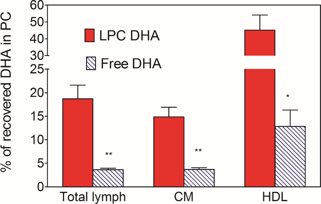
Fig. 3. Percent of DHA recovered in PC.
The PC and TAG from the lipids of total lymph, HDL, and CM were separated on aminopropyl columns, and the fatty acid composition was determined by GC/MS. The percent of DHA recovered in PC was calculated from this data. The PC fraction contained a small amount of DHA as PE (<10%), since these two lipids were not separated in this procedure. All the remaining DHA was in TAG. The results shown are mean ± SEM. (n= 10 total lymph for LPC-DHA, n=8 for total lymph for free DHA, n=5 each HDL and CM for both micelle).
* P<0.05; ** p<0.005 free DHA vs LPC-DHA (unpaired t test)

