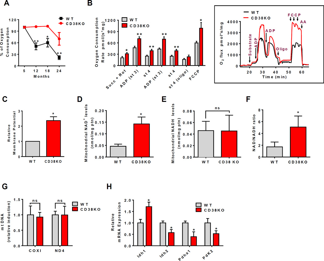
Figure 3. CD38 deficiency upregulates mitochondrial function and increases NAD+/NADH ratio in mitochondria.
Liver of WT and CD38KO 12 month old litter mate mice were used for the measurements below in B-H. All values are mean ± SEM.
(A) Percentage of oxygen consumption coupled to ATP synthesis in isolated mitochondria during aging in WT and CD38KO mice (n=4 animals for each age, *p<0.05, **p<0.01 versus 5 month old mice).
(B) Oxygen consumption rates in isolated mitochondria. The following drugs were added for the experimental profile: Succinate 10mM and rotenone 1µM (Succ+Rot), 0.15mM ADP, 1µg/mL oligomycin (Oligo), and 1µM FCCP (inset) (n=4, *p<0.05, **p<0.01 versus WT mice).
(C) Relative membrane potential in mitochondria (*p<0.05 versus WT mice).
(D–F) Total NAD+ levels (D), NADH levels (E), and the NAD+/NADH ratio (F) in isolated mitochondria (n=3, *p<0.05 versus WT mice).
(G) Relative mtDNA quantification of COX I and ND4 as mitochondrial-encoded genes normalized by GAPDH.
(H) Relative mRNA expression of glucose metabolism pathway enzymes (n=6, *p<0.05 versus WT mice).

