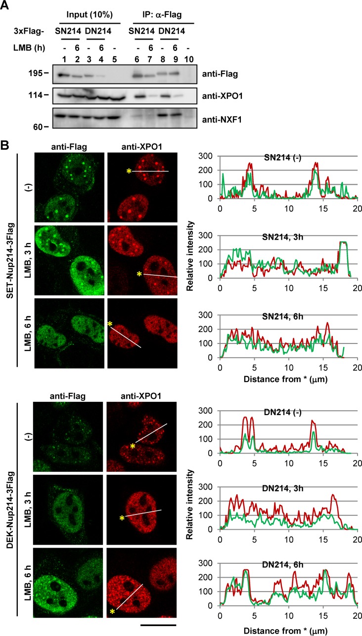
FIG 2.
Dependency of NES proteins on complex and dot formation by either SET-Nup214 or DEK-Nup214 with XPO1. (A) HeLa cells cultured in 35-mm dishes were transiently transfected with 1 μg of either pCAGGS-SET-Nup214-3Flag (SN214), DEK-Nup214-3Flag (DN214), or pCAGGS. At 2 days after transfection, cells were incubated in 5 ng/ml LMB (L-6100; LC Laboratories) for 6 h. After incubation, cells were collected and were subjected to IP assays using Flag M2 beads (lanes 6 to 10). Proteins in the input lysate and immunoprecipitated samples were separated by 6.5% SDS-PAGE, and Western blot analyses were performed using anti-Flag and anti-XPO1 antibodies as primary antibodies. Molecular weights (in thousands) of prestained markers are indicated on the left. (B) The protocol was the same as that for panel A. After cells were collected, IF analyses were performed. Anti-Flag M2 (1:1,000) and anti-XPO1 were used as primary antibodies. Bar, 20 μm. Graphs on the right represent the relative intensities of Flag-tagged protein (green) and XPO1 (red).

