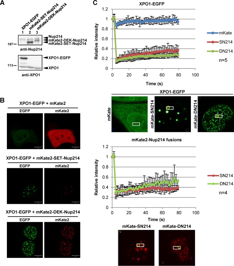
FIG 4.
Decreased mobility of XPO1. (A) HEK293T cells cultured in 35-mm dishes were transiently transfected with 1 μg pHCF1 (XPO1-EGFP expression vector), pmKate2C-SET-Nup214, or pmKate2C-DEK-Nup214. Samples were separated by 5% SDS-PAGE and were subjected to Western blot analyses using anti-Nup214 (dilution, 1:1,000) and anti-XPO1 antibodies. Molecular weights (in thousands) of prestained markers are indicated on the left. (B and C) HeLa cells were transfected with 1 μg pHCF1 and 1 μg either pmKate2C, pmKate2C-SET-Nup214, or pmKate2C-DEK-Nup214. (B) Typical localization patterns of XPO1-EGFP, mKate2, mKate2-SET-Nup214, and mKate2-DEK-Nup214 are shown. Bars, 10 μm. (C) Transfected cells were subjected to FRAP assays as described previously (82).

