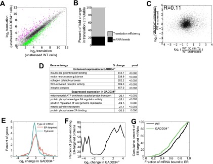
FIG 2.
Regulation of protein synthesis by GADD34 in unstressed MEFs. (A) Comparison of total levels of mRNA translation, analyzed by ribosome profiling, in unstressed WT and unstressed GADD34−/− MEFs (n = 2). The shaded area represents 5 standard deviations from the mean of results from WT biological replicates, with genes with significantly different results (at least 2-fold change and P < 0.05 by Student's t test [52]) highlighted in magenta (enhanced translation in the absence of GADD34) or green (suppressed translation in the absence of GADD34). (B) Percentages of changes in total translation in unstressed GADD34−/− cells relative to WT cells attributable to changes in mRNA levels and translation efficiency. Percentages were calculated as described in Materials and Methods. (C) Relationship between changes in translation in unstressed GADD34−/− cells relative to WT cells and changes in translation in WT cells after 30 min of induction of ER stress by the use of 1 μM Tg as reported previously (22). (D) Gene ontologies with changes in total translation in the absence of GADD34, with P values (p-val) determined by bootstrapping. (E) Histogram of differences in total translation between GADD34−/− and WT MEFs for mRNAs encoding ER-targeted proteins (containing signal sequence or transmembrane domain) (red) and cytosolic proteins (green). (F) Moving averages were calculated for the proportions of mRNAs encoding ER-targeted proteins as a function of GADD34-mediated changes in translation. Enrichment among the highly suppressed and highly enhanced genes highlights mRNAs encoding ER-targeted proteins as particularly sensitive to loss of GADD34. (G) Cumulative density plot representing the fraction of each mRNA encoding ER-targeted proteins associated with ER in WT and GADD34−/− cells.

