FIG 4.
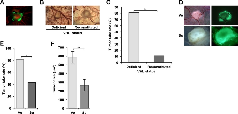
786-O cells form tumors on the chorioallantoic membrane (CAM) of chicken embryos in a VHL gene- and sunitinib-dependent manner. (A) Fluorescence image of a tumor generated from 786-O cells (labeled with GFP) implanted on CAM. Green, tumor cells; red, Alexa Fluor 555-dextran labeling blood vessels. (B) Bright-field images of tumors on CAM using 786-O reconstituted with empty vector (Deficient) or VHL gene (Reconstituted). (C) Tumor take rates of VHL gene-deficient and reconstituted 786-O cells. (D) Bright-field and fluorescence images of 786-O-derived tumors on CAM treated with vehicle (Ve) or sunitinib (Su). (E) Tumor take rates of 786-O cells treated with Ve or Su. (F) Area of 786-O tumors treated with Ve or Su. Data are means ± SEM (n = 11 for Ve and n = 6 for Su). *, P < 0.05; **, P < 0.01.

