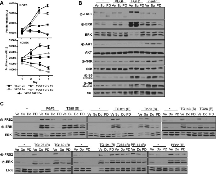
FIG 9.
FGFR activation by RCC. (A) Proliferation of HUVEC and HDMEC treated with Ve, Su (100 nM), or Do (500 nM) in the absence or presence of VEGF (100 ng/ml) or FGF2 (50 ng/ml) or both. Data are means ± SEM. n = 4. (B) Western blot analysis of MEFs pretreated with Ve, Su, Do, or PD and stimulated with VEGF (50 ng/ml), FGF2 (25 ng/ml), or insulin (300 nM). (C) Western blot analysis of MEFs pretreated with Ve, Su, Do, or PD and stimulated with conditioned media (CM) obtained from the indicated RCC cells. Su was used at 200 nM, Do at 1 μM, and PD at 100 nM for MEFs. (S), sunitinib sensitive; (R), sunitinib resistant; −, cells in the same base medium that was used to collect RCC-conditioned media.

