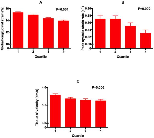
Figure 1. Relationship between quartiles of waist-hip-ratio and markers of systolic cardiac mechanics.
(A) global longitudinal strain, (B) peak systolic strain rate, and (C) tissue s’ velocity. Tissue velocities based on speckle-tracking echocardiography are lower than conventional tissue Doppler imaging tissue velocities. P-values are for the linear trend (calculated using linear mixed effects models).

