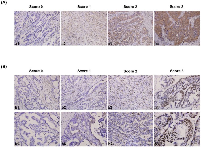
Figure 1.
Immunohistochemical staining for CDK5 and p27 expression in gastric cancerous tissue and the criteria for immunohistochemistry scoring. (A) Intensity score for CDK5 expression in gastric cancer tissue. a1 to a4: no staining (score 0), weak staining (score 1), moderate staining (score 2) and strong staining (score 3). The protein expression was considered low if the score was 1 or less and high if it was 2 or more. Scale bar = 100μm. (B) Distribution and intensity score for p27 expression in gastric cancer tissue. b1 to b4 (distribution score): ≤ 5% positive cells (score 0), 6% to 25% positive cells (score 1), 26% to 50% positive cells (score 2) and ≥ 51% positive cells (score 3). Scale bar = 100μm. b5 to b8 (intensity score): no staining (score 0), weak staining (score 1), moderate staining (score 2) and strong staining (score 3). The protein expression was considered low if the total score (distribution score + intensity score) was 3 or less and high if it was 4 or more. Scale bar = 50μm.

