
Figure 4. Demonstration of adhesions (pegs) between the detached hyaloid and retina in eyes with diabetic retinopathy on vitreous enhanced three-dimensional swept-source optical coherence tomography imaging.
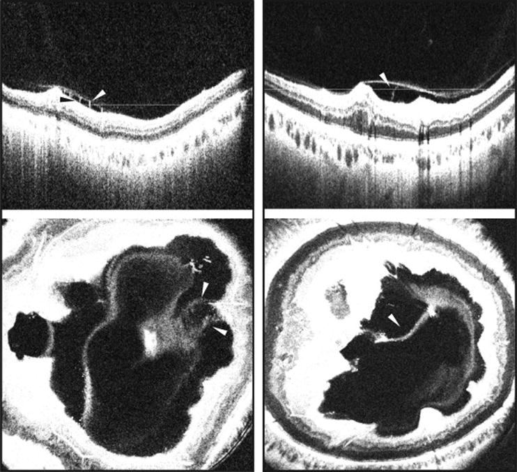
(Left Part) SS-OCT depicting a B-scan (top) and enface representation (bottom) of an eye with DME and severe NPDR showing adhesions (pegs) traversing the optically clear hypo-reflective space between the detached posterior hyaloid and the inner retinal surface. (Right Part) SS-OCT depicting a B-scan (top) and enface representation (bottom) of an eye with PDR showing adhesions (pegs) between the detached posterior hyaloid and retina. White and black arrowheads: Adhesions (pegs) between the detached posterior hyaloid and the inner retinal surface. White lines: location of the respective enface sections. SS-OCT, swept source optical coherence tomography; NPDR, non-proliferative diabetic retinopathy; PDR, proliferative diabetic retinopathy; DME, diabetic macular edema