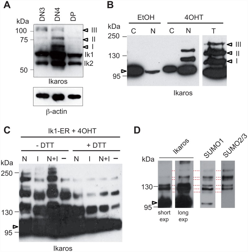
Fig 1. Ikaros is sumoylated in T cells.
(A) 5x10⁵ DN3, DN4 or DP cells were lysed in 1x sample buffer, subjected to SDS-PAGE (8%), and probed with an anti-Ikaros antibody. Arrowheads point to the modified fractions. (B) Cytosolic (C), nuclear (N) or total (T) fractions from 106 EtOH- or 4OHT-treated ILC87-IK1-ER cells were subjected to SDS-PAGE (10%) and analyzed with an anti-Ikaros antibody. (C) Western blot analysis (6% SDS-PAGE) with an anti-Ikaros antibody of total cell extracts from 2x10⁶ 4OHT-treated ILC87-IK1-ER cells that were lysed in the presence, absence or combination of N-ethylmaleimide (N), iodacetic acid (IAA, I) or 20 mM DTT, as indicated. (D) 4OHT-treated ILC87-Ik1-ER cells were lysed in buffer containing 1% NP-40 in the presence of NEM and IAA. Nuclear extracts from 50x106 cells were immunoprecipitated with an anti-ER antibody and analyzed on a NuPAGE Novex 3–8% Tris-Acetate gel with the indicated antibodies. In (B), (C) and (D), arrowheads point to unmodified Ik1-ER proteins.

