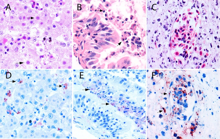
Fig 3. Microscopic lesions in amphibians naturally infected with CMTV-like ranavirus from the Netherlands.
These lesions illustrate the criteria considered in the double-blind semi-quantitative scoring system. (A) Hematoxylin and eosin (H&E)-stained liver of a Pelophylax kl.esculentus infected with CMTV-like ranavirus, black arrows indicate basophilic intracytoplasmic inclusion bodies in the hepatocytes. Original magnification ×400. (B) H&E–stained intestine of a Pelophylax kl.esculentus infected with CMTV-like ranavirus presenting with detachment and necrosis of enterocytes in the apical portion of the mucosal villi (black arrows). Original magnification ×400. (C) H&E-stained section of the intestinal submucosa with evident vascular damage characterized by perivascular edema and collections of karyorrhectic cell debris. Original magnification ×400. (D) Immunohistochemistry of a serial section from figure A using an anti-European catfish virus (ECV) polyclonal antibody, positive immunolabeling is observed in the cytoplasm of affected hepatocytes (black arrows). Original magnification ×400. (E) Immunohistochemistry of a serial section from figure B using ECV polyclonal antibody, positive immunolabeling is observed in numerous necrotic enterocytes exfoliated into the lumen (black arrows). Original magnification ×400. (F) Immunohistochemistry of a serial section from figure C using ECV polyclonal antibody, positive immunolabeling is present in the endothelial cell wall and in the cells scattered throughout the damaged submucosa (black arrows). Original magnification x400.

