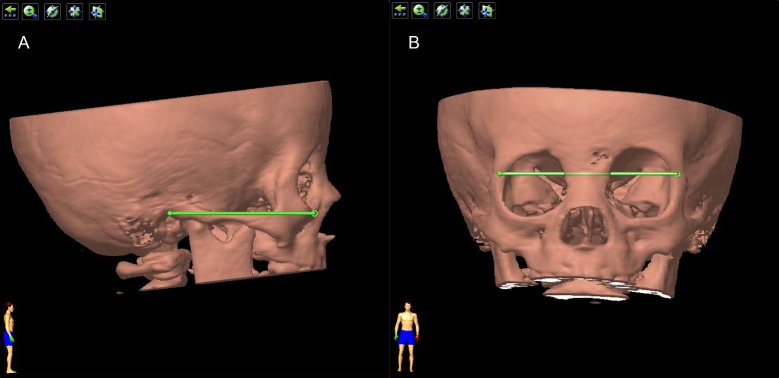
Fig 4. Standard position of the orbit.
(A) The ‘standard lateral position’. The 3D image of the orbit was adjusted until both mandibles coincide and the Reid’s base line was horizontal. The Reid’s base line was the green line drawn from the inferior margin of the orbit to the superior margin of the orifice of the external acoustic meatus. (B) The ‘standard frontal position’. The orbit was rotated by 90° from standard lateral potion, and then adjusted until the line drawn between two frontozygomatic sutures (the green line) was horizontal.

