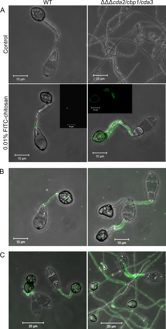
Fig 6. Fluorescently labelled chitosan is associated with the cell wall of the CDA deletion strains.
A) Rescue of appressorium development in the cda2/cbp1/cda3 deletion strain using exogenous, fluorescently-labelled chitosan, following a 16 hr incubation, showing clear localization to the cell wall of the germ tube and appressorium (single z-section insert). B) Rescue of appressorium development after a 2 hr incubation with FITC-chitosan, followed by washing and a further 16 hr incubation, showing germ tube-localized FITC-chitosan. C) Rescue of appressorium development after addition of FITC-chitosan at 16 hpi, and incubation for a further 8 hr. Scale bars: as shown in pictures.

