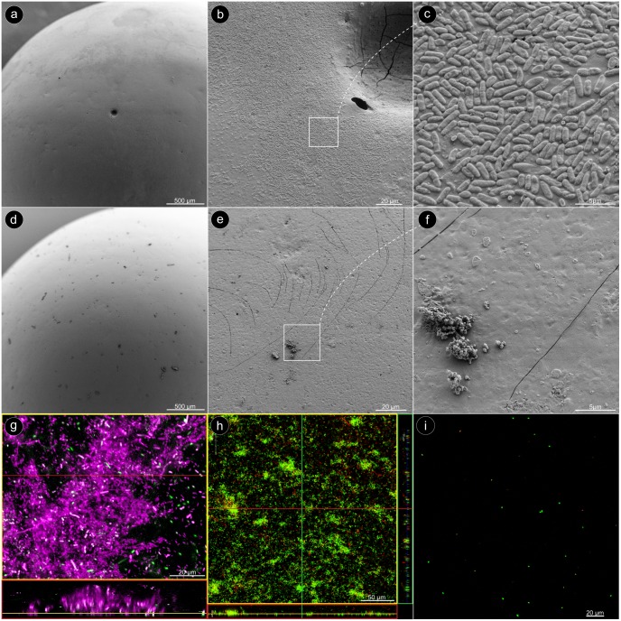
Fig 2. Microscopic characterization.
(a) SEM image: overview on a glass bead after 24 h cultivation with P. aeruginosa. (b) The bead surface is evenly covered with biofilm. (c) The bacteria are densely arranged in a monolayer. (d) Overview on a glass bead after the biofilm had been removed by sonication. (e, f) The bead surface is virtually empty, except for some residual debris. (g) CLSM image: The sugar-matrix of the P. aeruginosa biofilm was stained with Concanavalin A (assigned color: magenta), and the bacteria with Syto60 (assigned color: green). (h) LIVE/DEAD® staining of the biofilm on a glass bead. (i) LIVE/DEAD® staining after the biofilm had been removed from the bead by sonication.

