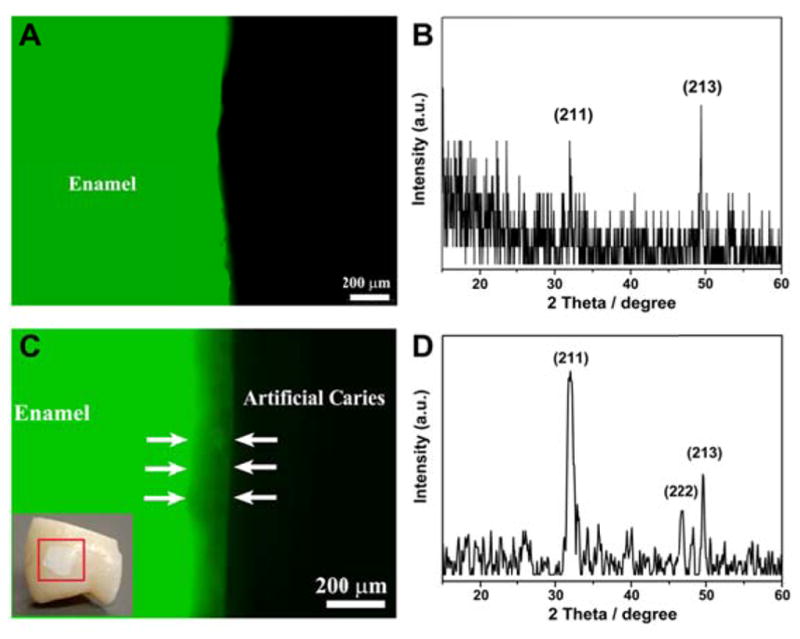
Figure 3.

Representative fluorescent images of cross-sections of (A) healthy enamel and (C) demineralized enamel. Inset: photograph of a demineralized enamel block showing a white spot lesion on the tooth sample (red square). XRD spectra of (B) healthy enamel surface and (D) enamel surface with the caries-like lesions.
