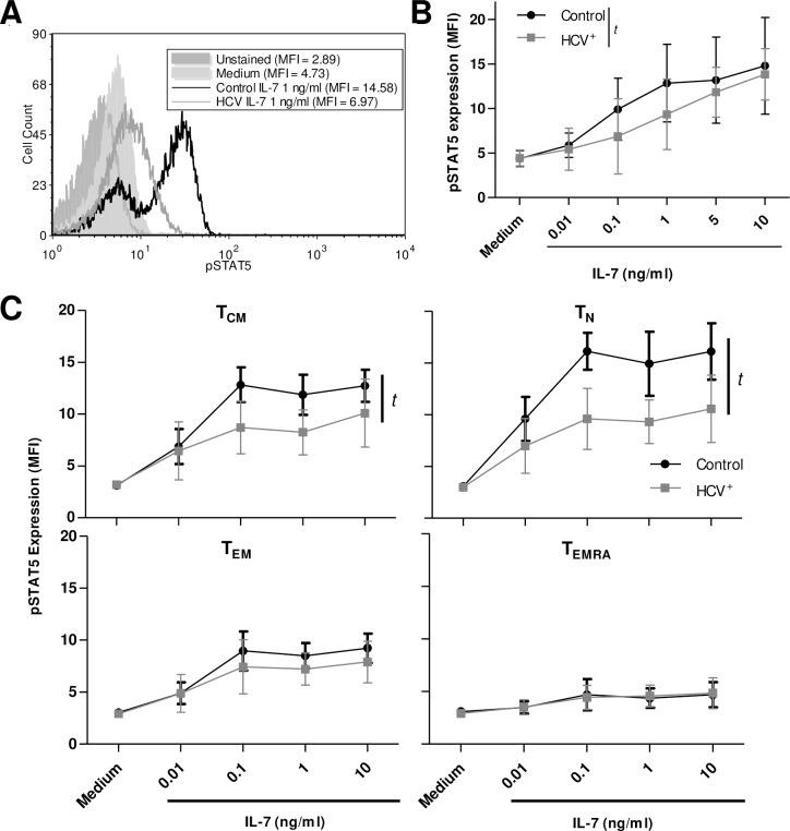
Fig 2. IL-7-induced signaling of blood-derived CD8+ T-cells is impaired in HCV infection.
(A) Phosphorylation of STAT5 was measured as mean fluorescence intensity (MFI) as shown in a representative histogram. (B) The expression of pSTAT5 was significantly increased by increasing concentrations of IL-7 (0.01–10 ng/ml) in blood-derived CD8+ T-cells from controls (p < 0.001, n = 10) or chronically infected HCV+ individuals (p = 0.003, n = 9) is summarized, as assessed by ANOVA, yet responses of the latter group were significantly less pronounced than controls (t: p = 0.005, non-linear regression analysis). (C) The expression of pSTAT5 in CD8+ T-cell subsets were distinguished by CD45RA and CCR7 expression, with significance in TCM and TN subsets (t: p<0.0001 for each subset, non-linear regression, control n = 7, HCV+ n = 5). Error bars in the graphs represent ± S.D.

