FIGURE 1.

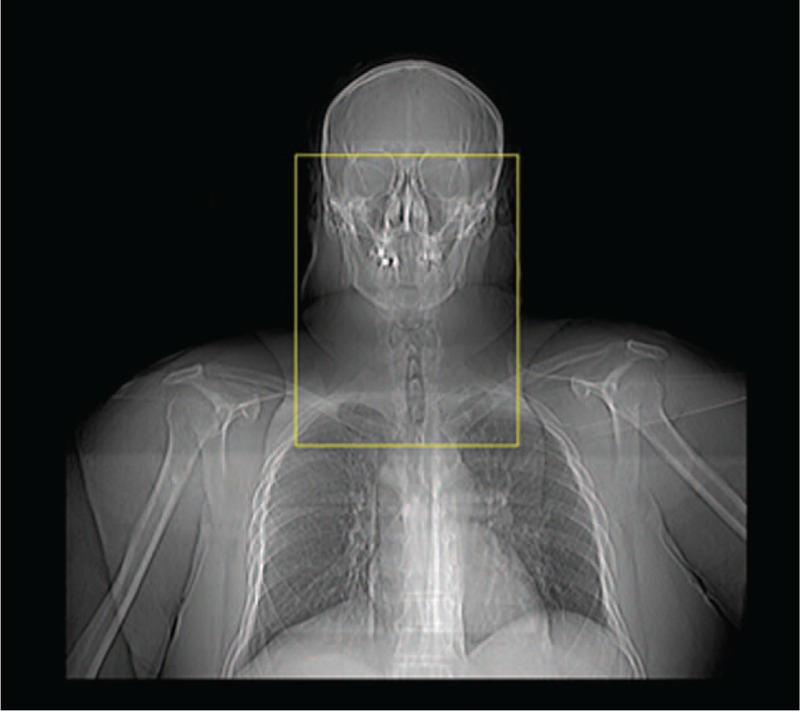
Computed tomography (CT) margins obtained from the scout view: from the roof of the bony orbits cranially, to the sternal angle of lewis inferiorly, and from the mid-clavicular interspace laterally.

Computed tomography (CT) margins obtained from the scout view: from the roof of the bony orbits cranially, to the sternal angle of lewis inferiorly, and from the mid-clavicular interspace laterally.