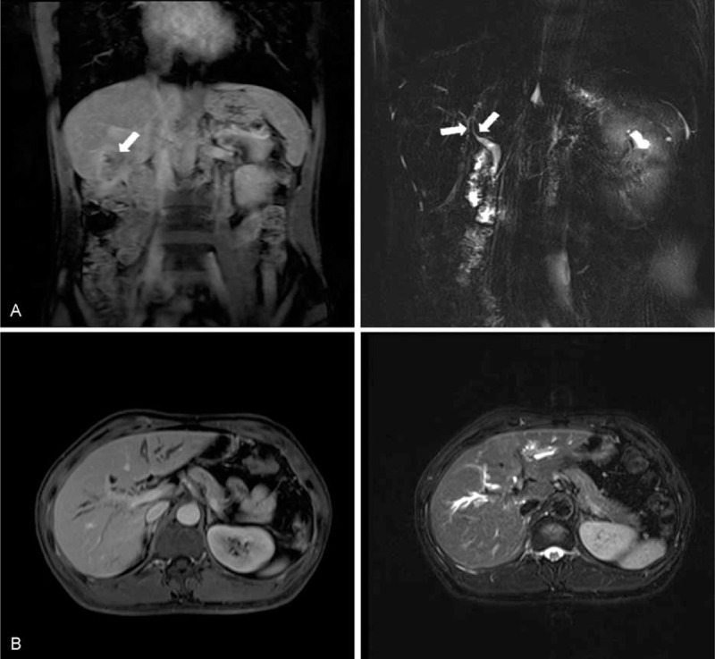
FIGURE 1.

Preoperative magnetic resonance cholangiopancreatography images. (A) Coronal reconstructed image revealed an irregular and abnormal signal intensity lesion (approximately 3.7 cm) in segment VI of liver mimicking malignancy (left), and the obstructed bile duct (right). Arrows show the stricture of the intrahepatic bile ducts caused by the cholangiocarcinoma. (B) Transversal sections on T1-weighted (left) and T2-weighted (right) images showed a slight enhancement of the common bile duct. Dilation and irregularity of biliary tracts were also obvious.
