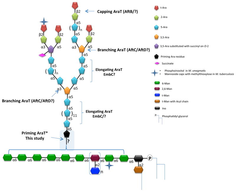
Figure 1. Structure of LAM.
The mannan core of LM and LAM is composed on average of 20–25 α–(1→6)-linked Manp residues occasionally substituted at C-2 by single Manp units in M. smegmatis and M. tuberculosis. Single Manp substitutions occur at C-3 in M. chelonae 25. Due to the findings reported herein, we have purposely left the details of the attachment of the arabinan to the mannan very general rather than the previously believed idea that it is attached at O-2 of one of the 6-linked mannosyl residues.

