Fig. 2.
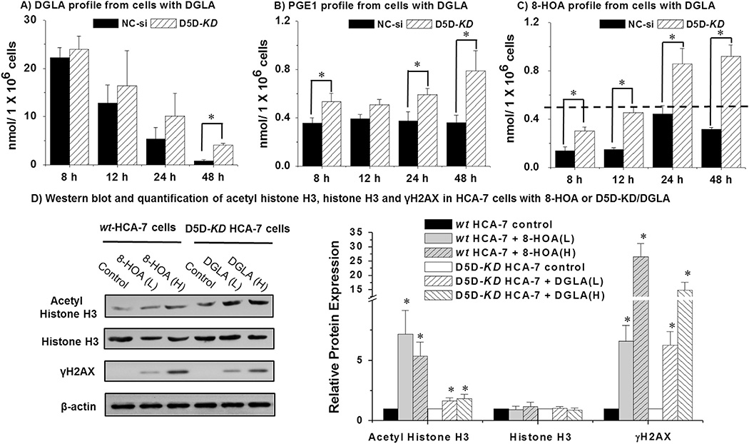
D5D-KD increased the levels of free DGLA, PGE1 and 8-HOA in HCA-7 colony 29 cells. (A) LC/MS quantification of DGLA from cell medium containing 1.0 × 10⁶ of control siRNA transfected or D5D-KD HCA-7 cells after DGLA treatment (100 µM); (B) LC/MS quantification of PGE1 from cell medium containing 1.0 × 10⁶ of control siRNA transfected or D5D-KD HCA-7 cells after DGLA treatment (100 µM); (C) GC/MS quantification of 8-HOA from cell medium containing 1.0 × 10⁶ of control siRNA transfected or D5D-KD HCA-7 cells after DGLA treatment (100 µM). Note: in the cell experiments without DGLA treatment, only trace amount of DGLA were detected within 48 h (≤ 0.03 respectively), while the formation of PGE1 and 8-HOA was under detect limit; (D) Western blots of acetyl-histone H3, histone H3 and γH2AX in HCA-7 cells treated with 8-HOA (10 and 25 µM) and D5D-KD HCA-7 cells treated by DGLA (50 and 100 µM). The relative ratios of different proteins to β-actin were normalized to 1 respectively; (*: significant difference vs. the corresponding controls with p < 0.05 from n ≥ 3).

