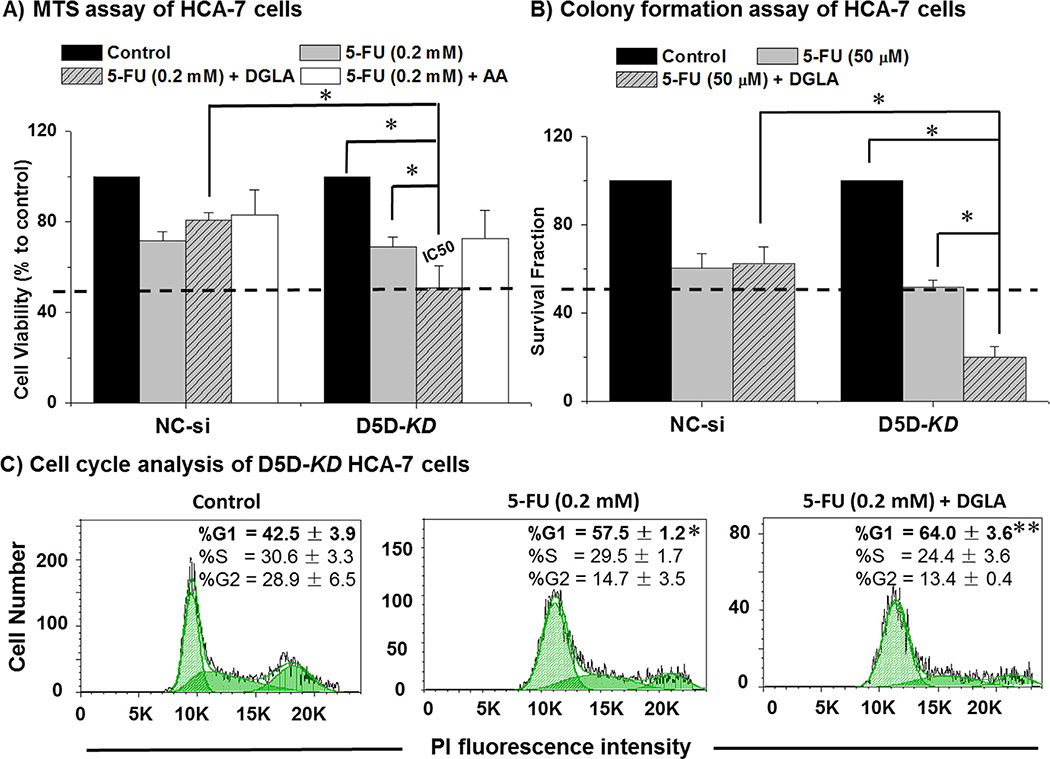
Fig. 3.
Enhanced 5-FU's efficacy by DGLA in D5D-KD HCA-7 colony 29 cells. (A) MTS assay for proliferation of HCA-7 cells (control siRNA transfected vs. D5D-KD) treated with 5-FU (0.2 mM), 5-FU + DGLA (100 µM) or 5-FU + AA (100 µM) for 48 h. The control siRNA transfected and D5D-KD cells without fatty acid and drug treatment were used as controls. The IC50 of 5-FU was improved to ~0.2 mM from 1.0 mM [2,49]. (B) Colony formation assay of HCA-7 cells (control siRNA transfected vs. D5D-KD) at 10 days with treatment of DGLA (100 µM), 5-FU (50 µM) or 5-FU + DGLA (100 µM) for 48 h. The control siRNA transfected and D5D-KD cells without fatty acid and drug treatment were used as controls. (*: significant difference with p < 0.05 from n ≥ 3). (C) Cell Cycle distribution was examed via flow cytometer afterD5D-KD HCA-7 cells were treated with 5-FU (0.2 mM), or 5-FU + DGLA (100 µM) for 48 h, followed by PI staining. At least 10,000 cells were counted for each sample. Cells treated with vehicle served as controls. (*: significant difference vs. control with p < 0.05; **: significant difference vs. 5-FU group with p < 0.05, n ≥ 3).

