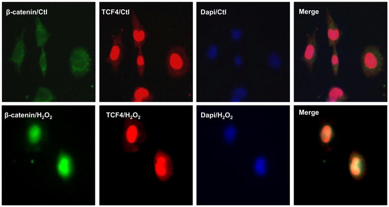
Figure 1. H2O2 induced β-catenin nuclear localization but TCF4 was not affected.
HL-1 cardiomyocytes were infected with β-catenin-GFP adenovirus for 24 hours, treated with 50 μM H2O2 for 1 hour, and observed by confocal microscopy. β-catenin-GFP adenovirus successfully infected the HL-1 cells and 50 μM H2O2 promoted β-catenin-GFP (green) accumulated in the nuclei. TCF4 were stained with Texas red (red). TCF4 accumulated in the nuclei at the baseline and did not change after 50 μM H2O2 treatment in HL-1 cells. n=3 batches of cells from 3 independent experiments carried on separate occasions for each group.

