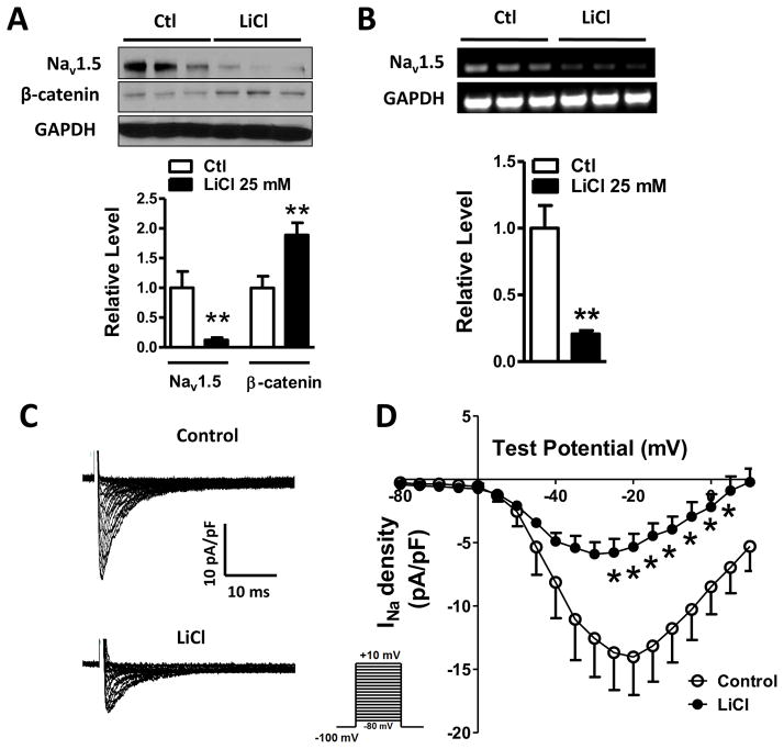
Figure 4. LiCl inhibited cardiac Na+ channel activity by suppressing NaV1.5 expression through activating Wnt/β-catenin signaling.
(A) Exposure of HL-1 cardiomyocytes to 25 mM LiCl led to a significant increase in β-catenin expression (p<0.01), compared to the control group (p<0.01), accompanied by a significant decrease of NaV1.5 protein (p<0.01). (B) RT-PCR was performed to detect the effect of 25 mM LiCl on NaV1.5 mRNA. NaV1.5 mRNA level was significantly decreased in HL-1 cells (p<0.01) after 25 mM LiCl treatment, compared to the control group. In A and B, Ctl: control; n=3 batches of cells from 3 independent experiments carried on separate occasions for each group. (C) INa currents were recorded in HL-1 cardiomyocytes with or without LiCl treatment. 25 mM LiCl decreased Na+ current densities HL-1 cells. (D) The current-voltage relationship curve of Na+ currents showed that the current densities obtained from peak currents divided by individual cell capacitances were significantly decreased from −25 mV to +5 mV in HL-1 cells (p<0.05), compared to the control group. In C and D, n=7 in the control group and n=7 in the LiCl group. In A, B and D, *p<0.05 and **p<0.01.

