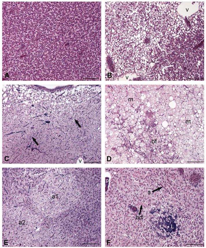
Figure 2.
Morphology of liver and hepatocellular alterations in Fundulus heteroclitus. A, normal parenchyma, note uniformity of hepatocytes and their staining. B-F altered liver. B, basophilic focus (bf); v, sections through veins. Due to the absence of overt hepatopancreas, venous profiles cannot be identified as portal (afferent) or hepatic (efferent). C, clear cell focus; arrows point to border of lesion; v, vein. D, mixed focus composed of basophilic focus (bf) and microvesicular vacuolation (m) surrounding the focus. E, clear cell adenomas (a1 and a2) are separated by a band of normal-appearing tissue. F, lymphocyte aggregate (L) in hepatic parenchyma near bile ductules (bd); s, sinusoid; venous profile at upper left of field shows portion of exocrine pancreatic tissue (p), likely indicating portal vein. All scale bars 10 μm. 20X, H&E.

