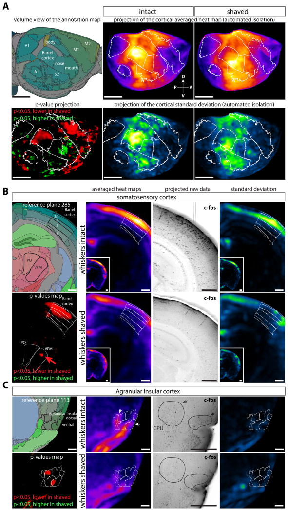
Figure 5. Brain regions differentially regulated by whiskers during an exploration task.
Mice had their whiskers shaved or left intact, and allowed to explore a new cage in the dark for 1hr, then brains where harvested and activity was probed by c-Fos immunolabeling (n=5 (shaved group), n=3 (control group)). A Lateral projections of the reference annotation, averaged density maps, p-value maps and standard deviation maps (automated isolation of the neo-cortex). The caudo-medial part of the barrel shows a decreased activity in the shaved group. B Coronal projection at the level of the barrel cortex and VPM thalamic relay, showing decreased activity in the whisker projection field (arrow in the VPM p-value map). C Decreased activity in the Agranular Insula (anterior part) in the shaved group. Abbreviations: CPU: Caudoputamen nucleus, Po: Posterior nucleus of the Thalamus, VPM: VentroPostero Medial nucleus of the thalamus. Scale bars are 2mm (panel a) and 500μm (panels b and c).

