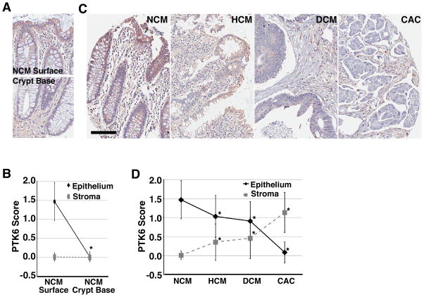
Figure 5. PTK6 expression decreases in epithelial cells during tumor progression.
Representative images from a tissue microarray with immunohistochemical staining for PTK6 in normal colonic mucosa (NCM) (A), and in NCM, hyperplastic (HCM), and dysplastic (DCM) colonic mucosa, and colonic adenocarcinoma (CAC) (C) (size bar = 100 μm). (B, D) PTK6 staining intensity was scored on a 0, 1+, 2+ scale for epithelial and stromal cells for all tissue samples.

