Fig. 1.

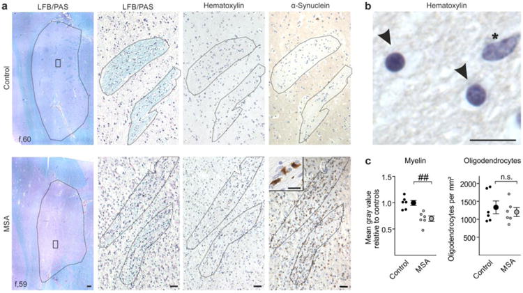
Putaminal myelin loss in multiple system atrophy (MSA) without alterations in oligodendrocyte density. a Representative Luxol Fast Blue/Periodic Acid Schiff (LFB/PAS) stained images (left) depict the putamen (encircled) of a control (upper panel) and a MSA patient (lower panel). The putamen of the MSA patient showed a reduced Luxol Fast Blue staining intensity reflecting severe loss of myelin. Scale bar: 500μm. Boxed areas within the left images are magnified showing LFB/PAS-, hematoxylin-, and α-synuclein-stained striae. Note abundant α-synuclein-positive inclusions in the putamen of the MSA patient (magnified as insert). Scale bars: 50μm, insert 10μm. b Oligodendrocytes (arrowheads; round-shaped, dense chromatin) were distinguished by morphology from astrocytes (asterisk; larger and more elongated in size, loose chromatin) in the hematoxylin stained-sections. Scale bar: 10μm. c While myelin was significantly reduced in MSA patients (gray dots) compared to controls (black dots), oligodendrocyte density was unaltered (n = 6). Data are shown as mean ± standard error of mean. T-test: ##p < 0.01, n.s. (not significant) p > 0.05. f = female.
