Fig. 7.

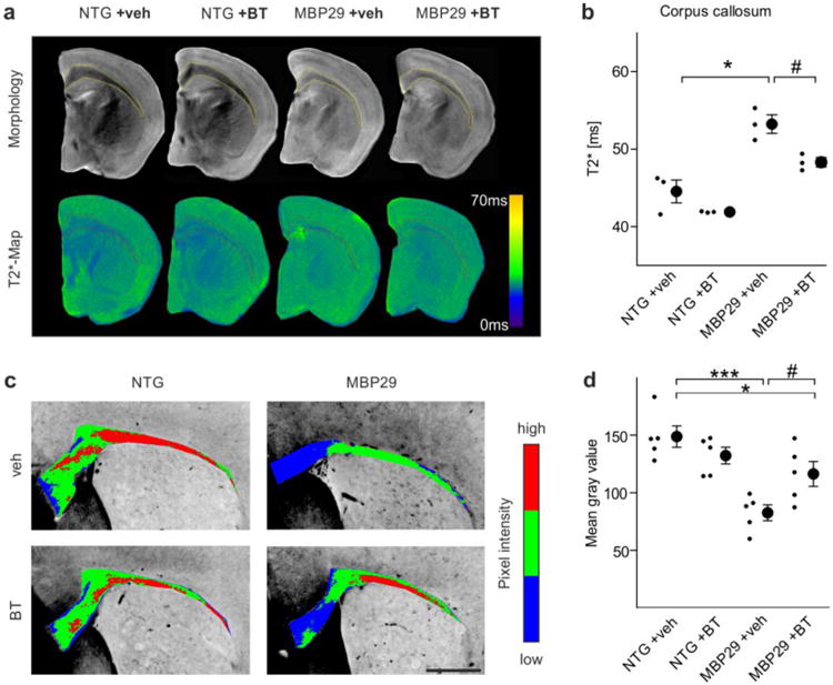
Amelioration of the myelin deficit in mice with oligodendrocytic overexpression of α-synuclein (MBP29) upon benztropine treatment. a Callosal myelin levels of non-transgenic (NTG) and MBP29 mice treated with vehicle (veh) or benztropine (BT, 2mg/kg) were analyzed using magnetic resonance imaging. Upper panel illustrates morphology as determined by T2-weighted magnetic resonance imaging, whereas lower panel depicts T2*-weighted magnetic resonance imaging (shown as color-coded maps), which was used for myelin quantification. b MBP29 mice (n = 3) showed a reduction in myelin levels detected by increased T2*-relaxation times within the corpus callosum. Benztropine attenuated the myelin deficit in MBP29 confirmed by a reduction of T2*. c Representative pictures of a Luxol Fast Blue staining (color-coded for signal intensity: blue low, green intermediate, red high intensity), confirming the effects of α-synuclein overexpression and benztropine treatment on myelination. Scale bar: 500μm. d MBP29 mice (n = 5) exhibited a reduction in Luxol Fast Blue intensity, which was attenuated, but not completely restored by benztropine. Data are shown as mean ± standard error of mean. ANOVA: *p < 0.05, ***p < 0.001. T-test: #p < 0.05.
